SPS Grewal,Manpreet Brar,Mansi Sharma,Mangat Ram Dogra,Dilraj S Grewal
OCT Atlas
OCT Atlas
YOU SAVE £8.40
- Condition: Brand new
- UK Delivery times: Usually arrives within 2 - 3 working days
- UK Shipping: Fee starts at £2.39. Subject to product weight & dimension
Bulk ordering. Want 15 or more copies? Get a personalised quote and bigger discounts. Learn more about bulk orders.
Couldn't load pickup availability
- More about OCT Atlas
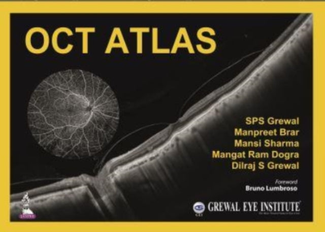
Optical coherence tomography (OCT) is a non-invasive imaging test that uses light waves to take cross-sectional pictures of the retina. This atlas provides OCT images to help with the identification, diagnosis, and treatment of common retinal and anterior segment disorders. It is divided into two sections, with images illustrating the normal fundus and numerous different retinal disorders. Section two covers anterior segment disorders, including corneal dystrophies, ocular surface disorders, keratoconus, glaucoma, and trauma.
Format: Hardback
Length: 220 pages
Publication date: 29 November 2021
Publisher: Jaypee Brothers Medical Publishers
Optical coherence tomography (OCT) is a non-invasive imaging technique that employs light waves to capture cross-sectional images of the retina, the light-sensitive tissue lining the back of the eye. This innovative method is widely recognized as a crucial tool for the diagnosis, assessment, and follow-up of various retinal diseases and glaucoma.
This comprehensive atlas serves as a valuable resource for ophthalmologists and trainees, offering a collection of OCT images that aid in the identification, diagnosis, and treatment of common retinal and anterior segment disorders. The images in this atlas have been meticulously compiled from the authors' personal collections using advanced Plex Elite and Cirrus 6000 technology. Additionally, fundus angiography images are included to provide further insight into related pathologies.
The book is organized into two sections. The first section focuses on the normal fundus, presenting a series of images that depict the various structures and components of the retina in a healthy state. This section serves as a foundation for understanding the subsequent chapters, which delve into the diverse range of retinal disorders.
The second section encompasses anterior segment disorders, beginning with images of the normal cornea. It then proceeds to illustrate a wide array of conditions, including corneal dystrophies, ocular surface disorders, keratoconus, glaucoma, and trauma. Each section is accompanied by a multitude of images, each accompanied by brief descriptive text.
By utilizing the images and descriptive text provided in this atlas, ophthalmologists and trainees can gain a deeper understanding of the various retinal and anterior segment disorders, enabling them to provide accurate diagnoses, effective treatment plans, and optimal patient care.
Weight: 2825g
Dimension: 330 x 241 (mm)
ISBN-13: 9789354650925
This item can be found in:
UK and International shipping information
UK and International shipping information
UK Delivery and returns information:
- Delivery within 2 - 3 days when ordering in the UK.
- Shipping fee for UK customers from £2.39. Fully tracked shipping service available.
- Returns policy: Return within 30 days of receipt for full refund.
International deliveries:
Shulph Ink now ships to Australia, Belgium, Canada, France, Germany, Ireland, Italy, India, Luxembourg Saudi Arabia, Singapore, Spain, Netherlands, New Zealand, United Arab Emirates, United States of America.
- Delivery times: within 5 - 10 days for international orders.
- Shipping fee: charges vary for overseas orders. Only tracked services are available for most international orders. Some countries have untracked shipping options.
- Customs charges: If ordering to addresses outside the United Kingdom, you may or may not incur additional customs and duties fees during local delivery.


