
Donald E.Thrall,Ian D.Robertson
Atlas of Normal Radiographic Anatomy and Anatomic Variants in the Dog and Cat
Atlas of Normal Radiographic Anatomy and Anatomic Variants in the Dog and Cat
YOU SAVE £23.38
- Condition: Brand new
- UK Delivery times: Usually arrives within 2 - 3 working days
- UK Shipping: Fee starts at £3.89. Subject to product weight & dimension
Bulk ordering. Want 15 or more copies? Get a personalised quote and bigger discounts. Learn more about bulk orders.
Couldn't load pickup availability
- More about Atlas of Normal Radiographic Anatomy and Anatomic Variants in the Dog and Cat
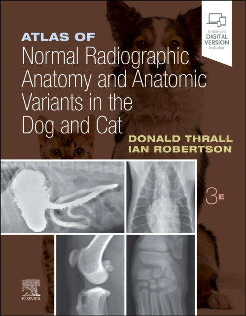
The 3rd Edition of Atlas of Normal Radiographic Anatomy and Anatomic Variants in the Dog and Cat is a comprehensive atlas that offers detailed images of normal and non-standard subjects, along with demonstrations of proper technique and image interpretations. It provides a wider range of normal subjects, including overweight and underweight pets, and animals with breed-specific variations. Updated with new high-contrast images, descriptive text, and explanatory legends, the atlas helps practitioners make accurate diagnoses and achieve successful treatment outcomes.
Format: Hardback
Length: 320 pages
Publication date: 23 September 2022
Publisher: Elsevier - Health Sciences Division
This highly visual, comprehensive atlas offers an in-depth exploration of both normal and non-standard subjects in dog and cat anatomy, providing a valuable resource for accurate diagnoses and successful treatment outcomes. With an extensive collection of new high-contrast images, Atlas of Normal Radiographic Anatomy and Anatomic Variants in the Dog and Cat, 3rd Edition, offers a detailed examination of various body parts and structures. Expert authors Donald E. Thrall and Ian D. Robertson have expanded the coverage of normal subjects, showcasing standard dogs and cats as well as non-standard subjects such as overweight and underweight pets and animals with breed-specific variations.
Each body part is accompanied by a descriptive text that explains the underlying radiographic principles, enabling readers to appreciate variations of normal based on a solid understanding of basic radiographic concepts. The atlas also includes updated descriptive text and explanatory legends that accompany all images, further enhancing the understanding of anatomical structures.
High-quality digital images provide excellent contrast resolution and enhance visibility of normal structures, aiding in accurate diagnoses. In-depth coverage of patient positioning and radiographic exposure guidelines assists practitioners in achieving optimal results.
The third edition has been expanded to include additional coverage of neonatal and juvenile subjects, providing valuable radiographic examples for these critical stages of development. Furthermore, the atlas now includes expanded material on the normal appearance of various special procedures performed in private practice, such as barium esophagram, barium gastrointestinal study, and positive contrast cystogram.
Additionally, the atlas features radiographic images of normal or standard prototypical animals, allowing readers to compare their anatomy with the standard reference model. This comprehensive resource is an invaluable tool for veterinarians, veterinary students, and anyone interested in studying dog and cat anatomy.
Weight: 1108g
Dimension: 224 x 284 x 19 (mm)
ISBN-13: 9780323796156
Edition number: 3 ed
UK and International shipping information
UK and International shipping information
We deliver throughout the United Kingdom and to 128 countries and territories worldwide, including the United States, Australia, Canada, Germany, Spain and France.